


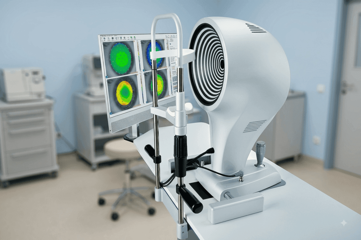
Corneal Topography: What It Measures, Clinical Applications & How to Choose a Corneal Topographer
Corneal topography is an important diagnostic technology used to evaluate the shape, curvature, and optical characteristics of the cornea. By generating detailed maps of the corneal surface, a corneal topographer can provide information that may not be captured through conventional keratometry alone.
Modern corneal topography systems are used in ophthalmic clinics, refractive surgery centres, contact lens practices, diagnostic centres, and eye hospitals for applications including corneal assessment, keratoconus screening, refractive evaluation, contact lens fitting, and pre- and post-operative analysis.
For healthcare organisations selecting ophthalmic diagnostic equipment, understanding what a corneal topographer measures, how the technology works, and which features matter can help make procurement decisions more effectively.
What Is Corneal Topography?
Corneal topography is a non-contact imaging and measurement technique used to create a detailed map of the corneal surface.
The cornea is not perfectly spherical. Its curvature can vary from the centre toward the periphery, and irregularities in its shape can affect how light is refracted through the eye.
A conventional keratometer primarily evaluates selected areas of the cornea and provides measurements of the principal meridians. Corneal topography can provide a much more detailed representation of corneal curvature across a broader area.
The resulting maps can help ophthalmic professionals evaluate patterns of corneal steepening, flattening, asymmetry, elevation, and other characteristics depending on the technology used.
For clinics building a complete diagnostic workflow, corneal topography is therefore often considered alongside other corneal measurement technologies such as keratometry and pachymetry.
Explore Medsprings' broader Ophthalmic Equipment portfolio for additional diagnostic and measurement solutions.
How Does Corneal Topography Work?
Different corneal topographers use different imaging and measurement principles.
One of the most established approaches is Placido disk-based topography. In this method, concentric illuminated rings are projected onto the cornea. The reflected pattern is captured by a camera and analysed using computer algorithms to calculate characteristics of the corneal surface.
The shape and spacing of the reflected rings change according to the curvature of the cornea. The resulting information can then be converted into colour-coded maps and numerical measurements.
Modern systems may combine Placido disk imaging with additional technologies such as optical tomography, Scheimpflug imaging, or other elevation-based approaches.
This distinction is important when comparing corneal topographers because different technologies can provide different types of information about the cornea.
What Does a Corneal Topographer Measure?
Depending on the technology and software configuration, corneal topography systems can provide several types of measurements and maps.
1. Corneal Curvature
One of the primary purposes of corneal topography is to evaluate variations in corneal curvature.
Curvature information can be presented across multiple points of the corneal surface rather than being limited to a small number of measurements.
This can help identify areas that are relatively steep, flat, or irregular.
2. Corneal Refractive Power
Corneal topographers can calculate the refractive power of different areas of the cornea.
This information can be useful when evaluating the optical characteristics of the corneal surface and assessing irregular patterns.
3. Axial or Sagittal Maps
An axial, or sagittal, map represents corneal curvature using a colour-coded format.
These maps are commonly used to understand the overall distribution of corneal power and curvature.
4. Tangential Maps
Tangential maps provide another way of analysing local curvature changes.
They can be particularly useful when examining localized variations in corneal curvature and surface irregularity.
5. Elevation Maps
Elevation mapping compares measured corneal surfaces against a reference surface.
Depending on the imaging technology, systems can provide anterior and posterior elevation information.
This can be particularly useful in more advanced corneal assessment where understanding three-dimensional corneal shape is important.
6. Pachymetry Information
Some advanced corneal imaging systems can provide corneal thickness or pachymetry maps.
Corneal thickness is an important parameter in several areas of ophthalmic assessment and can complement curvature and elevation data.
A dedicated Pachymeter can also be used when precise corneal thickness measurement is required.
Corneal Topography vs Keratometry
Corneal topography and keratometry are related, but they are not the same.
A keratometer measures the curvature of selected principal meridians of the cornea. It is widely used for applications such as keratometry and contact lens-related measurements.
Corneal topography provides a more detailed map of corneal curvature across a larger area.
In simple terms:
| Feature | Keratometer | Corneal Topographer |
|---|---|---|
| Primary purpose | Corneal curvature measurement | Detailed corneal surface mapping |
| Measurement coverage | Selected areas/meridians | Broader corneal surface |
| Output | Numerical curvature measurements | Colour-coded maps and numerical data |
| Local irregularity assessment | More limited | More detailed |
| Corneal mapping | Limited | Yes |
| Advanced elevation analysis | Depends on device | Available on advanced systems |
| Keratoconus assessment | Limited | Common application |
| Contact lens assessment | Yes | Yes, with appropriate software |
Medsprings also offers a Manual Keratometer for conventional corneal curvature measurement.
For clinics, the choice between a keratometer and a corneal topographer should depend on the intended applications rather than simply comparing specifications.
Corneal Topography vs Corneal Tomography
The terms corneal topography and corneal tomography are sometimes used interchangeably, but they describe different approaches to corneal imaging.
Traditional Placido-based corneal topography primarily evaluates the anterior corneal surface through reflected ring patterns.
Tomography-based systems can provide information about the three-dimensional structure of the cornea, including anterior and posterior surfaces and corneal thickness, depending on the technology.
Modern systems may combine different imaging technologies to provide a more comprehensive assessment.
Therefore, when selecting a system, clinics should consider whether they primarily need corneal surface mapping or require broader three-dimensional corneal analysis.
Clinical Applications of Corneal Topography
Corneal topography has applications across several areas of ophthalmic practice.
1. Keratoconus Screening
One of the important applications of corneal topography is the evaluation of corneal patterns associated with keratoconus and other corneal ectatic disorders.
Early keratoconus can present with subtle changes that may not be obvious through routine examination alone. Detailed corneal mapping can provide additional information about curvature and shape.
Corneal topography and tomography are widely used in the evaluation of keratoconus and suspected ectatic disorders.
A corneal topographer should not, however, be viewed as a standalone replacement for comprehensive clinical examination. Results should be interpreted by appropriately qualified eye-care professionals alongside other clinical findings.
2. Refractive Surgery Assessment
Corneal topography can form part of the pre-operative assessment for refractive procedures.
Detailed mapping may help clinicians evaluate corneal shape and identify patterns that require additional consideration before surgery.
Topographic assessment may also be used for post-operative evaluation to examine changes in corneal curvature.
3. Contact Lens Fitting
Corneal shape varies significantly between individuals.
Detailed corneal mapping can therefore support the assessment and fitting of specialty contact lenses, particularly where conventional measurements may not provide enough information.
Some modern corneal topographers include dedicated contact lens simulation or fitting software.
4. Assessment of Irregular Astigmatism
Corneal topography can help identify irregular patterns in corneal curvature.
This may be relevant in patients with corneal scars, previous surgery, keratoconus, or other corneal conditions that can alter the normal corneal surface.
5. Pre- and Post-Operative Evaluation
Corneal mapping may be used before and after ophthalmic procedures to document corneal shape and evaluate changes over time.
The exact role depends on the procedure and the clinical protocol used by the ophthalmologist.
6. Corneal Wavefront Assessment
Advanced systems may provide corneal wavefront or aberrometry-related analysis.
This can provide additional information about optical characteristics of the corneal surface and may be useful in specialised ophthalmic and refractive applications.
What Are the Different Types of Corneal Topography Technology?
When choosing a corneal topographer, it is useful to understand the main technologies available.
Placido Disk-Based Topography
Placido disk systems project concentric rings onto the cornea and analyse their reflection.
They are particularly useful for mapping anterior corneal curvature and are widely used in clinical corneal assessment.
Scheimpflug-Based Imaging
Scheimpflug imaging uses a rotating or angled camera system to capture cross-sectional images of the cornea.
Depending on the system, this can provide information about anterior and posterior corneal surfaces, elevation, and pachymetry.
Optical Coherence Tomography
Anterior segment OCT can provide high-resolution cross-sectional imaging of ocular structures.
Some modern corneal imaging systems incorporate OCT or related optical tomography technologies to expand the type of information available.
Combined Technologies
Advanced systems may combine more than one measurement principle.
For example, a system can combine Placido disk-based surface analysis with tomography or OCT-based imaging.
This can provide a broader set of measurements within a single diagnostic platform.
Key Features to Consider When Choosing a Corneal Topographer
Selecting a corneal topography machine should begin with the clinical requirements of the facility.
1. Measurement Technology
First, determine which measurement principle the system uses.
Consider whether the clinic needs:
-
Placido disk corneal mapping
-
Elevation-based imaging
-
Scheimpflug imaging
-
OCT-based imaging
-
A combination of technologies
A system that combines technologies may offer a broader diagnostic dataset, depending on the intended clinical applications.
2. Corneal Coverage
The area of the cornea covered during measurement is an important specification.
A larger measurement area can provide more information about the peripheral cornea and overall corneal shape.
However, coverage should always be evaluated alongside measurement quality and the intended clinical application.
3. Acquisition Speed
Fast acquisition can be useful in busy ophthalmic clinics and diagnostic centres.
It can help reduce examination time and make the equipment more practical for higher patient volumes.
4. Repeatability and Accuracy
Measurement repeatability is an important consideration when comparing corneal topographers.
Procurement teams should review the manufacturer's stated accuracy and repeatability specifications and consider how the system performs under the intended clinical workflow.
5. Map Types
A useful corneal topographer should provide map types appropriate to the clinic's requirements.
Depending on the system, these may include:
-
Axial/sagittal maps
-
Tangential maps
-
Elevation maps
-
Refraction maps
-
Pachymetry maps
More map types can be useful, but only when the clinical team can interpret and use the additional information effectively.
6. Keratoconus Screening Software
If the system will be used for corneal ectasia assessment, review the available keratoconus screening and analysis software.
Automated indices and screening tools can help organise information, but they should support rather than replace professional clinical interpretation.
7. Contact Lens Simulation
For clinics providing specialty contact lens services, contact lens simulation can be a valuable feature.
This may be particularly relevant for practices working with complex corneal shapes.
8. Ease of Use
A sophisticated diagnostic system should still fit into the clinic's workflow.
Consider:
-
Patient positioning
-
Automated alignment
-
Auto-capture
-
User interface
-
Report generation
-
Data storage
-
Multi-map display
-
Training requirements
Ease of operation can influence throughput and consistency in day-to-day use.
9. Display and Software
The software environment is just as important as the hardware.
A good system should allow clinicians to review maps clearly, compare measurements, examine numerical data, and generate reports appropriate to their workflow.
10. Supplier and After-Sales Support
For hospitals, clinics, distributors, and international buyers, procurement should also consider the supplier behind the equipment.
Important factors can include:
-
Product availability
-
Documentation
-
Technical support
-
Installation assistance
-
Training
-
Spare parts and service support
-
International shipping
-
Response time
-
Long-term supply relationship
For international procurement, working with an ophthalmic supply partner that understands export requirements can simplify sourcing and communication.
Medsprings Corneal Topographer
Medsprings offers a non-contact corneal topography system designed for ophthalmic and refractive applications.
The system combines Placido Disk Projection with Anterior Optical Tomography/OCT and provides multiple measurement and analysis capabilities.
According to the product specifications, the system includes:
-
22–32 concentric Placido rings
-
Less than 1 second per scan acquisition
-
Corneal coverage of up to 10–12 mm
-
Dioptric power range of 10.0 D to 100.0 D
-
Accuracy/repeatability of ±0.1 D to ±0.5 D
-
Axial/sagittal maps
-
Tangential maps
-
Anterior and posterior elevation maps
-
Refraction maps
-
Pachymetry maps
-
Keratoconus screening software
-
Corneal wavefront analysis
-
Contact lens fitting simulation
-
Multi-map comparative display
The system is also designed for non-contact measurement and includes applications related to early keratoconus and ectasia screening, corneal wavefront analysis, and specialty contact lens assessment.
You can view the complete Medsprings Corneal Topographer product specifications and submit a product enquiry.
Who Can Benefit From a Corneal Topographer?
Corneal topography can be relevant for several types of ophthalmic facilities.
Eye Hospitals
Hospitals may use corneal mapping as part of broader diagnostic and surgical assessment workflows.
Ophthalmology Clinics
Clinics can use corneal topography for corneal assessment, refractive evaluation, contact lens-related applications, and monitoring.
Refractive Surgery Centres
Detailed corneal mapping can be useful in pre-operative assessment and post-operative evaluation for appropriate patients.
Specialty Contact Lens Practices
Corneal shape data can support specialty contact lens assessment and fitting.
Diagnostic Centres
Standalone diagnostic facilities may require corneal topography as part of a broader ophthalmic diagnostic equipment portfolio.
Medical Distributors
Distributors sourcing ophthalmic diagnostic equipment should evaluate not only the product specifications but also documentation, supply continuity, technical support, and the supplier's ability to support their target market.
Corneal Topography in a Modern Ophthalmic Equipment Setup
Corneal topography is generally one part of a broader ophthalmic diagnostic workflow.
Depending on the clinical environment, a facility may also require:
-
Keratometers
-
Pachymeters
-
Tonometry equipment
-
Auto refractometers
-
Biometers
-
Ophthalmoscopes
-
Slit lamps
-
Vision testing equipment
-
Corneal imaging systems
Medsprings' Ophthalmic Equipment portfolio includes corneal measurement and keratometry solutions alongside tonometry, biometry, lens measurement, ocular examination, and vision and refraction equipment.
For a broader procurement overview, see Essential Ophthalmic Diagnostic Equipment for Modern Eye Care.
You can also read How to Choose Ophthalmic Equipment for an Eye Clinic for a wider equipment-selection framework.
How to Choose the Right Corneal Topographer for Your Clinic
A practical selection process can be divided into five steps.
Step 1: Define the Clinical Application
Identify why the equipment is being purchased.
Is the primary requirement:
-
General corneal mapping?
-
Keratoconus screening?
-
Refractive surgery assessment?
-
Specialty contact lens fitting?
-
Corneal wavefront analysis?
-
Research or advanced diagnostic imaging?
The answer will determine which features are actually necessary.
Step 2: Determine the Required Measurement Technology
Compare Placido-based, Scheimpflug, OCT, and combined systems according to the information required by the clinical team.
Step 3: Compare Measurement and Software Features
Review:
-
Corneal coverage
-
Acquisition speed
-
Repeatability
-
Map types
-
Elevation data
-
Pachymetry
-
Keratoconus analysis
-
Contact lens simulation
-
Wavefront analysis
-
Report generation
Step 4: Evaluate Workflow
Consider how easily the equipment can be integrated into the existing clinic.
Fast acquisition, intuitive software, automated alignment, and clear reporting can be important for facilities with higher patient volumes.
Step 5: Evaluate the Supplier
Finally, consider the supply relationship.
For international buyers, hospitals, distributors, and healthcare procurement teams, factors such as product documentation, communication, availability, shipping support, and after-sales assistance can be just as important as the technical specifications.
Frequently Asked Questions About Corneal Topography
What is corneal topography?
Corneal topography is a non-contact diagnostic technique that maps the shape and curvature of the cornea. It can generate colour-coded maps and numerical information about corneal surface characteristics.
What is a corneal topographer used for?
A corneal topographer can be used for corneal mapping, keratoconus screening, refractive assessment, contact lens fitting, evaluation of irregular astigmatism, and pre- and post-operative corneal assessment.
Can corneal topography detect keratoconus?
Corneal topography can help identify corneal patterns associated with keratoconus and other ectatic disorders. However, results should be interpreted by a qualified eye-care professional together with other clinical findings.
What is the difference between corneal topography and keratometry?
Keratometry measures corneal curvature at selected principal meridians, while corneal topography provides a more detailed map of corneal curvature across a broader surface.
Is corneal topography the same as corneal tomography?
No. Traditional corneal topography primarily maps the anterior corneal surface, while tomography can provide three-dimensional information about the cornea, including anterior and posterior surfaces and thickness, depending on the imaging technology.
What is a Placido disk corneal topographer?
A Placido disk corneal topographer projects concentric rings onto the cornea and analyses their reflected pattern to calculate corneal surface characteristics.
What should I look for when buying a corneal topographer?
Important factors include measurement technology, corneal coverage, acquisition speed, accuracy and repeatability, available map types, keratoconus screening tools, contact lens simulation, software, ease of use, and supplier support.
Is corneal topography useful for contact lens fitting?
Yes. Corneal mapping can provide detailed information about corneal shape that may be useful for contact lens assessment and specialty lens fitting. Systems with contact lens simulation can provide additional functionality.
What other equipment is used for corneal measurement?
Depending on the clinical requirement, corneal measurement may also involve keratometers and pachymeters. Medsprings offers a Manual Keratometer and Pachymeter alongside its corneal topography system.
Conclusion
Corneal topography has become an important part of modern corneal assessment because it provides detailed information about corneal shape and curvature beyond conventional single-point or meridional measurements.
From keratoconus screening and refractive assessment to specialty contact lens fitting and corneal analysis, the technology can support a wide range of ophthalmic applications.
When selecting a corneal topographer, healthcare organisations should look beyond the headline specification. Measurement technology, corneal coverage, map types, acquisition speed, software capabilities, workflow, and supplier support should all be considered.
For hospitals, ophthalmology clinics, diagnostic centres, refractive practices, and international medical distributors looking for ophthalmic diagnostic equipment, Medsprings provides corneal topography alongside a broader portfolio of ophthalmic equipment and corneal measurement solutions.